What I've been doing for the past couple of days
Microinjecting and snapping photos ...
Here are some weird things that I've been seeing (or as they say IMAGING) in the nucleus. I wont explain much ... so view it as nano-art.
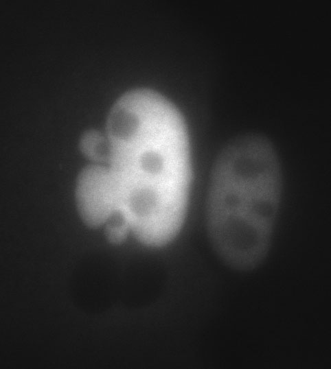
First up are two nuclei microinjected with Fluorescein coupled 70-KiloDalton Dextran (basically a large fluorescent molecule). The injected fluorophore redistributes throughout, but can't escape, the nucleus. Why can't it leave? It's too big. Notice the darker splotches within the nuclei, these are dense zones are called nucleoli, and are centers for ribosome assembly. Look at the multi-lobular nuclei on the left, how can a nuclei be deformed in such a manner? The answer is totally unknown.
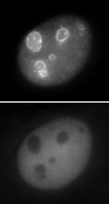
OK next up is a coinjection experiment. Two pictures of the same cell. Top picture is of RNA, stained with a red fluorophore, bottom picture is fluorescein (green) 70-kD dextran (see previous paragraph). Notice the cool looking dots of RNA that SURROUND THE NUCLEOLI - very weird. Notice how these dots are regularly spaced.
Again how or why these dots/granules/bodies are formed - totally unknown. If anyone ever tells you that we understand biology or cells - they obviously haven't been looking very hard.
So much to look at, so little time (to blog) ...
I'll post pictures of exploded cells next ...
posted by apalazzo at 11:14 AM
![]()
![]()


<< Home